|
| Details about selected cell |
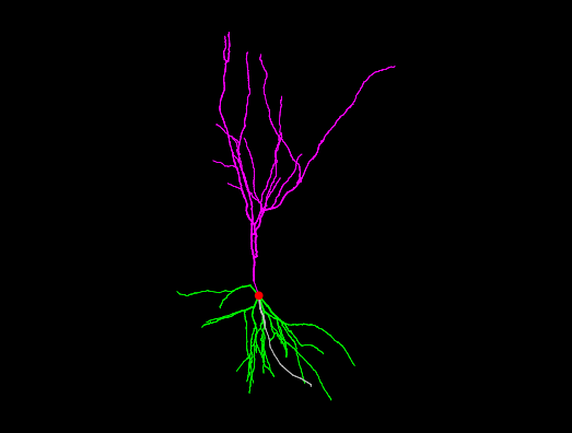
| NeuroMorpho.Org ID : | NMO_165878 |
| Cell Name : | PRZ-REMSD16_1 |
| Archive Name : | Mallick |
| Species Name : | rat |
| Strain : | Wistar |
| Structural Domains : | Dendrites, Soma, Axon |
| Physical Integrity : | Dendrites Moderate, Axon Incomplete |
| Morphological Attributes : | No Diameter, 3D, Angles |
| Min Age : | 6.0 months
|
| Max Age : | 6.0 months
|
| Gender : |
Male
|
| Min Weight : | 220 grams
|
| Max Weight : | 260 grams
|
| Development : | adult |
| Primary Brain Region : | hippocampus |
| Secondary Brain Region : | CA3 |
| Tertiary Brain Region : | Not reported |
| Primary Cell Class : | principal cell |
| Secondary Cell Class : | pyramidal |
| Tertiary Cell Class : | Not reported |
| Original Format : | Neurolucida.dat |
| Experiment Protocol : | in vitro |
| Experimental Condition : | recovered post-REMSD + Prazosin (alpha1 noradrenergic alpha1 receptor antagonist) treated |
| Staining Method : | Golgi-Cox |
| Slicing Direction : | coronal |
| Slice Thickness : | 250
μm |
| Tissue Shrinkage : |
Reported 11%
Not corrected
|
| Objective Type : | oil |
| Magnification : | 100x |
| Reconstruction Method : | Neurolucida |
| Date of Deposition : | 2021-08-04 |
| Date of Upload : | 2021-10-15 |
| Persistence Vector : | PRZ-REMSD16_1.pvec |
| Note : | This reconstruction was obtained from the recovered post-REMSD treated with Prazosin (alpha1 noradrenergic alpha1 receptor antagonist) group. The rats are maintained on small platforms surrounded by water so that they do not get enough space to relax completely to enjoy REMS and thus, are deprived of REMS. The recovery (REC) rats were first REMS deprived for 6 days and then kept for recovery for 3 days in their home cages. REMSD received intraperitoneal (IP) injection of alpha1 antagonist, prazosin (PRZ; 2 mg/kg) daily between 10 and 11 a.m. during 3rd to 6th day of REMSD, that is, during the last 4 days of REMSD (PRZ+REMSD). |
|
|
| Reference Article |
| Related Article Reference : | Rapid eye movement sleep deprivation impairs neuronal plasticity and reduces hippocampal neuronal arborization in male albino rats: Noradrenaline is involved in the process. |
|
|
|
|
| Measurements |
| Soma Surface : |
1587.79 μm2 |
| Number of Stems : |
6 |
| Number of Bifurcations : |
36 |
| Number of Branches : |
78 |
| Overall Width : |
204.85 μm |
| Overall Height : |
384.54 μm |
| Overall Depth : |
170.3 μm |
| Average Diameter : |
1.19 μm |
| Total Length : |
4254.56 μm |
| Total Surface : |
17613.7 μm2 |
| Total Volume : |
7641.43 μm3 |
| Max Euclidean Distance : |
323.29 μm |
| Max Path Distance : |
451.3 μm |
| Max Branch Order : |
5 |
| Average Contraction : |
0.85 |
| Total Fragmentation : |
1822 |
| Partition Asymmetry : |
0.38 |
| Average Rall's Ratio : |
1.18 |
| Average Bifurcation Angle Local : |
73.26° |
| Average Bifurcation Angle Remote : |
51.32° |
| Fractal Dimension : |
1.05 |
|
|
|
|
|
 0
0