|
| Details about selected cell |
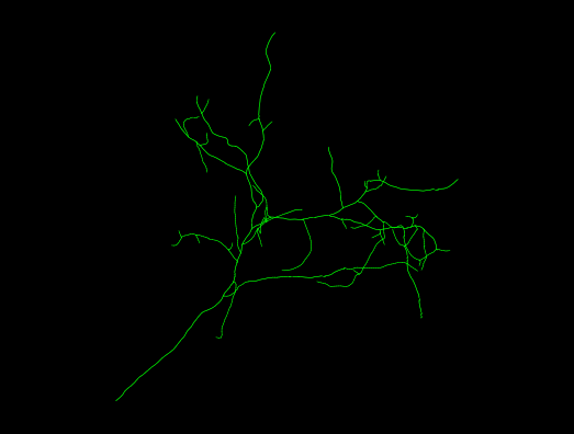
| NeuroMorpho.Org ID : | NMO_161891 |
| Cell Name : | Live1-19-2019_7-02-28_PM_denoised |
| Archive Name : | Ruthazer |
| Species Name : | Xenopus laevis |
| Strain : | Albino |
| Structural Domains : | Dendrites, No Soma, No Axon |
| Physical Integrity : | Dendrites Moderate |
| Morphological Attributes : | No Diameter, 3D, Angles |
| Min Age : | 42.0 days
|
| Max Age : | 44.0 days
|
| Gender : |
Not reported
|
| Min Weight : | Not reported
|
| Max Weight : | Not reported
|
| Development : | tadpole |
| Primary Brain Region : | mesencephalon |
| Secondary Brain Region : | tectum |
| Tertiary Brain Region : | optic |
| Primary Cell Class : | principal cell |
| Secondary Cell Class : | Not reported |
| Tertiary Cell Class : | Not reported |
| Original Format : | Imaris.imx |
| Experiment Protocol : | in vivo |
| Experimental Condition : | Control |
| Staining Method : | enhanced green fluorescent protein |
| Slicing Direction : | Not applicable |
| Slice Thickness : | Not applicable
|
| Tissue Shrinkage : |
Not reported
|
| Objective Type : | water |
| Magnification : | 60x |
| Reconstruction Method : | Imaris |
| Date of Deposition : | 2021-01-20 |
| Date of Upload : | 2021-08-06 |
| Persistence Vector : | Live1-19-2019_7-02-28_PM_denoised.pvec |
| Note : | Albino tadpoles at stage 44-46 according to the criteria of Nieuwkoop and Faber (1956) were used for electroporation. Animals were anesthetized by immersion in MS222 (0.02% in 0.1 x MBS-H). DNA solution containing various concentrations of either pEGFP-N1 or a mixture of pCAG-Cre with 1 micrograms/microliter. pCALNL-GFP and/or pCALNL-DsRed suspended in distilled water with a small amount of fast green dye for visualization was pressure injected in the brain ventricle with a glass micropipette. Reconstructions were obtained 5 days after the cre Mediated single-cell electroporation |
|
|
| Reference Article |
| Related Article Reference : | A Simple and Efficient Method for Visualizing Individual Cells in vivo by Cre-Mediated Single-Cell Labeling by Electroporation (CREMSCLE). |
|
|
|
|
| Measurements |
| Soma Surface : |
N/A |
| Number of Stems : |
1 |
| Number of Bifurcations : |
38 |
| Number of Branches : |
77 |
| Overall Width : |
56.09 μm |
| Overall Height : |
58.07 μm |
| Overall Depth : |
10.35 μm |
| Average Diameter : |
0.81 μm |
| Total Length : |
518.45 μm |
| Total Surface : |
1312.14 μm2 |
| Total Volume : |
264.27 μm3 |
| Max Euclidean Distance : |
82.97 μm |
| Max Path Distance : |
102.88 μm |
| Max Branch Order : |
14 |
| Average Contraction : |
0.93 |
| Total Fragmentation : |
1769 |
| Partition Asymmetry : |
0.51 |
| Average Rall's Ratio : |
2 |
| Average Bifurcation Angle Local : |
49.66° |
| Average Bifurcation Angle Remote : |
81.83° |
| Fractal Dimension : |
1.03 |
|
|
|
|
|
 0
0