|
| Details about selected cell |
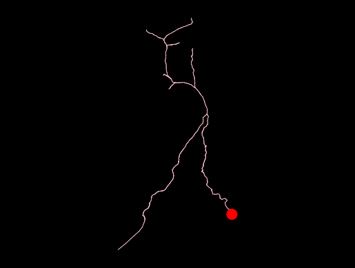
| NeuroMorpho.Org ID : | NMO_296355 |
| Cell Name : | L_neck_MN_D_bundle_N_10680 |
| Archive Name : | Kuan_Phelps |
| Species Name : | drosophila melanogaster |
| Strain : | y,w/w1118; +; P{VT025718-Gal4}attP2/P{pBI-UASC-3xMYC-sbAPEX2-dlg-S97}18 |
| Structural Domains : | Neurites, Soma |
| Physical Integrity : | Neurites Moderate |
| Morphological Attributes : | No Diameter, 3D, Angles |
| Min Age : | 1.0 day
|
| Max Age : | 2.0 days
|
| Gender : |
Female
|
| Min Weight : | Not reported
|
| Max Weight : | Not reported
|
| Development : | adult |
| Primary Brain Region : | peripheral nervous system |
| Secondary Brain Region : | ventral nerve cord |
| Tertiary Brain Region : | prothoracic neuromere (T1), left, dorsal, neck connective |
| Primary Cell Class : | principal cell |
| Secondary Cell Class : | Motoneuron |
| Tertiary Cell Class : | Not reported |
| Original Format : | Catmaid.swc |
| Experiment Protocol : | in vitro |
| Experimental Condition : | Control |
| Staining Method : | Osmium tetroxide, Potassium ferrocyanide, Thiocarbohydrazide, Uranyl acetate, Lead aspartate |
| Slicing Direction : | Not applicable |
| Slice Thickness : | Not applicable
|
| Tissue Shrinkage : |
Not reported
|
| Objective Type : | electron microscopy |
| Magnification : | Not reported
|
| Reconstruction Method : | Catmaid |
| Date of Deposition : | 2022-09-29 |
| Date of Upload : | 2025-02-26 |
| Persistence Vector : | L_neck_MN_D_bundle_N_10680.pvec |
| Note : | Leg MNs originate from 15 developmental lineages, and neurons from the same lineage have their primary neurites bundled together. Consistent with this, the EM-reconstructed front leg MN primary neurites appeared spatially clustered, forming 18 distinct bundles within the left and right T1 neuromeres. This reconstruction belongs to the D bundle. |
|
|
| Reference Article |
| Related Article Reference : | Reconstruction of motor control circuits in adult Drosophila using automated transmission electron microscopy. |
|
|
|
|
| Measurements |
| Soma Surface : |
1465.75 μm2 |
| Number of Stems : |
1 |
| Number of Bifurcations : |
6 |
| Number of Branches : |
13 |
| Overall Width : |
28.56 μm |
| Overall Height : |
199.39 μm |
| Overall Depth : |
95.96 μm |
| Average Diameter : |
0.72 μm |
| Total Length : |
551.54 μm |
| Total Surface : |
1175.04 μm2 |
| Total Volume : |
258.9 μm3 |
| Max Euclidean Distance : |
184.02 μm |
| Max Path Distance : |
314.85 μm |
| Max Branch Order : |
6 |
| Average Contraction : |
0.89 |
| Total Fragmentation : |
756 |
| Partition Asymmetry : |
0.83 |
| Average Rall's Ratio : |
1.85 |
| Average Bifurcation Angle Local : |
61.32° |
| Average Bifurcation Angle Remote : |
84.68° |
| Fractal Dimension : |
1.04 |
|
|
|
|
|
 0
0