|
| Details about selected cell |
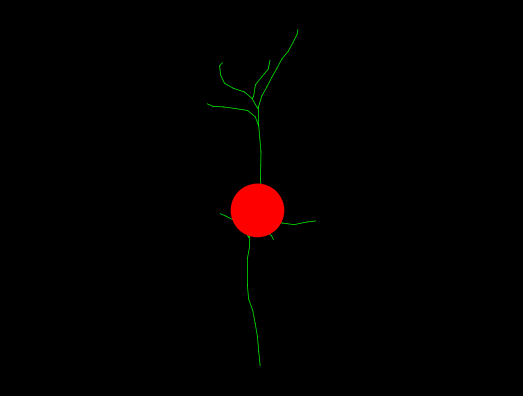
| NeuroMorpho.Org ID : | NMO_146741 |
| Cell Name : | GFP_15_bottom_1 |
| Archive Name : | Firestein |
| Species Name : | human |
| Strain : | Not applicable |
| Structural Domains : | Dendrites, Soma, No Axon |
| Physical Integrity : | Dendrites Complete |
| Morphological Attributes : | No Diameter, 2D, Angles |
| Min Age : | Not reported
|
| Max Age : | Not reported
|
| Gender : |
Not reported
|
| Min Weight : | Not reported
|
| Max Weight : | Not reported
|
| Development : | not reported |
| Primary Brain Region : | Not reported |
| Secondary Brain Region : | Not reported |
| Tertiary Brain Region : | Not reported |
| Primary Cell Class : | principal cell |
| Secondary Cell Class : | induced pluripotent stem cell-(iPSC)-derived |
| Tertiary Cell Class : | microtubule-associated protein 2 (MAP2) positive |
| Original Format : | NeuronJ.swc |
| Experiment Protocol : | culture |
| Experimental Condition : | D-serine treatment |
| Staining Method : | enhanced green fluorescent protein, immunostaining |
| Slicing Direction : | Not applicable |
| Slice Thickness : | Not applicable
|
| Tissue Shrinkage : |
Not reported
|
| Objective Type : | dry |
| Magnification : | 20x |
| Reconstruction Method : | NeuronJ |
| Date of Deposition : | 2020-11-16 |
| Date of Upload : | 2021-01-05 |
| Persistence Vector : | GFP_15_bottom_1.pvec |
| Note : | We transfected human neurons with constructs encoding GFP. The hiPSC-derived neurons were treated on DD19 by adding vehicle to the medium containing dimethyl sulfoxide (DMSO). The final concentration of DMSO in treatments was 0.1%. To maximally activate NMDAR glycine binding sites, neurons were treated for 48 h, from DD19-21, with 10 microM D-serine. Neuronal reconstructions were obtained at 21 days post-differentiation (DD21). |
|
|
| Reference Article |
| Related Article Reference : | Characterization hiPSC-derived neural progenitor cells and neurons to investigate the role of NOS1AP isoforms in human neuron dendritogenesis. |
|
|
|
|
| Measurements |
| Soma Surface : |
20.98 μm2 |
| Number of Stems : |
5 |
| Number of Bifurcations : |
3 |
| Number of Branches : |
11 |
| Overall Width : |
4.61 μm |
| Overall Height : |
15.24 μm |
| Overall Depth : |
0 μm |
| Average Diameter : |
0.3 μm |
| Total Length : |
33.21 μm |
| Total Surface : |
31.3 μm2 |
| Total Volume : |
2.35 μm3 |
| Max Euclidean Distance : |
8.97 μm |
| Max Path Distance : |
9.28 μm |
| Max Branch Order : |
3 |
| Average Contraction : |
0.91 |
| Total Fragmentation : |
62 |
| Partition Asymmetry : |
0.67 |
| Average Rall's Ratio : |
2 |
| Average Bifurcation Angle Local : |
88.94° |
| Average Bifurcation Angle Remote : |
65.39° |
| Fractal Dimension : |
1.04 |
|
|
|
|
|
 0
0