|
| Details about selected cell |
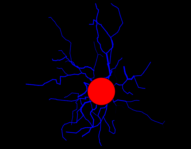
| NeuroMorpho.Org ID : | NMO_78231 |
| Cell Name : | DG-inner-mol-02_3 |
| Archive Name : | Papageorgiou_Kann |
| Species Name : | rat |
| Strain : | Wistar |
| Structural Domains : | Processes, Soma |
| Physical Integrity : | Processes Moderate |
| Morphological Attributes : | Diameter, 2D, Angles |
| Min Age : | 29.0 days
|
| Max Age : | 31.0 days
|
| Gender : |
Male
|
| Min Weight : | 115 grams
|
| Max Weight : | 130 grams
|
| Development : | young adult |
| Primary Brain Region : | hippocampus |
| Secondary Brain Region : | dentate gyrus |
| Tertiary Brain Region : | granule layer, inner |
| Primary Cell Class : | Glia |
| Secondary Cell Class : | microglia |
| Tertiary Cell Class : | Iba1-positive |
| Original Format : | Neurolucida.dat |
| Experiment Protocol : | ex vivo |
| Experimental Condition : | Control |
| Staining Method : | immunostaining |
| Slicing Direction : | horizontal |
| Slice Thickness : | 450
μm |
| Tissue Shrinkage : |
Not reported
|
| Objective Type : | dry |
| Magnification : | 40x |
| Reconstruction Method : | Neurolucida |
| Date of Deposition : | 2017-06-18 |
| Date of Upload : | 2017-11-28 |
| Persistence Vector : | DG-inner-mol-02_3.pvec |
|
|
| Reference Article |
| Related Article Reference : | Widespread activation of microglial cells in the hippocampus of chronic epileptic rats correlates only partially with neurodegeneration. |
|
|
|
|
| Measurements |
| Soma Surface : |
522.82 μm2 |
| Number of Stems : |
12 |
| Number of Bifurcations : |
21 |
| Number of Branches : |
54 |
| Overall Width : |
56.5 μm |
| Overall Height : |
51.7 μm |
| Overall Depth : |
1.22 μm |
| Average Diameter : |
0.85 μm |
| Total Length : |
639.55 μm |
| Total Surface : |
1706.55 μm2 |
| Total Volume : |
413.13 μm3 |
| Max Euclidean Distance : |
42.2 μm |
| Max Path Distance : |
54.79 μm |
| Max Branch Order : |
4 |
| Average Contraction : |
0.88 |
| Total Fragmentation : |
450 |
| Partition Asymmetry : |
0.25 |
| Average Rall's Ratio : |
1.47 |
| Average Bifurcation Angle Local : |
86.36° |
| Average Bifurcation Angle Remote : |
73.37° |
| Fractal Dimension : |
1.05 |
|
|
|
|
|
 0
0