|
| Details about selected cell |
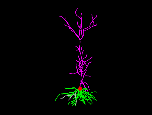
| NeuroMorpho.Org ID : | NMO_160932 |
| Cell Name : | Casp6_N73T_Neuron_6 |
| Archive Name : | Sjostrom |
| Species Name : | mouse |
| Strain : | C57BL/6 |
| Structural Domains : | Dendrites, Soma, Axon |
| Physical Integrity : | Dendrites & Axon Moderate |
| Morphological Attributes : | No Diameter, 3D, Angles |
| Min Age : | 11.0 days
|
| Max Age : | 16.0 days
|
| Gender : |
Male/Female
|
| Min Weight : | Not reported
|
| Max Weight : | Not reported
|
| Development : | young |
| Primary Brain Region : | hippocampus |
| Secondary Brain Region : | CA1 |
| Tertiary Brain Region : | pyramidal layer |
| Primary Cell Class : | principal cell |
| Secondary Cell Class : | pyramidal |
| Tertiary Cell Class : | Not reported |
| Original Format : | Neuromantic.swc |
| Experiment Protocol : | in vitro |
| Experimental Condition : | recombinant human caspase-6 N73T |
| Staining Method : | Alexa Fluor 594 |
| Slicing Direction : | custom |
| Slice Thickness : | 300
μm |
| Tissue Shrinkage : |
Not reported
|
| Objective Type : | water |
| Magnification : | 40x |
| Reconstruction Method : | Neuromantic |
| Date of Deposition : | 2021-07-20 |
| Date of Upload : | 2021-07-30 |
| Persistence Vector : | Casp6_N73T_Neuron_6.pvec |
| Note : | Recombinant human caspase-6 N73T was activated in Stennicke buffer at 37 Celsius degrees for 15 min and diluted to 10 pg/10 microL in internal solution with 30-60 micromolar Alexa 594 or 40-80 micromolar Alexa 488, and osmolarity double checked to be around 310 mOsm. These reconstructions were done from 2-photon laser-scanning microscopy image stacks, which means compartment diameters may appear larger than they are. These reconstructions may therefore not be suitable for multicompartmental computer simulations (Blackman et al., Frontiers in Synaptic Neuroscience 2013, DOI: 10.3389/fnsyn.2013.00011). |
|
|
| Reference Article |
| Related Article Reference : | Rare CASP6N73T variant associated with hippocampal volume exhibits decreased proteolytic activity, synaptic transmission defect, and neurodegeneration. |
|
|
|
|
| Measurements |
| Soma Surface : |
942.73 μm2 |
| Number of Stems : |
7 |
| Number of Bifurcations : |
77 |
| Number of Branches : |
161 |
| Overall Width : |
443.86 μm |
| Overall Height : |
1035.51 μm |
| Overall Depth : |
189.45 μm |
| Average Diameter : |
3.31 μm |
| Total Length : |
12401.4 μm |
| Total Surface : |
128751 μm2 |
| Total Volume : |
121547 μm3 |
| Max Euclidean Distance : |
960.74 μm |
| Max Path Distance : |
1085.43 μm |
| Max Branch Order : |
26 |
| Average Contraction : |
0.94 |
| Total Fragmentation : |
2165 |
| Partition Asymmetry : |
0.55 |
| Average Rall's Ratio : |
1.95 |
| Average Bifurcation Angle Local : |
69.15° |
| Average Bifurcation Angle Remote : |
54.91° |
| Fractal Dimension : |
1.02 |
|
|
|
|
|
 0
0