|
| Details about selected cell |
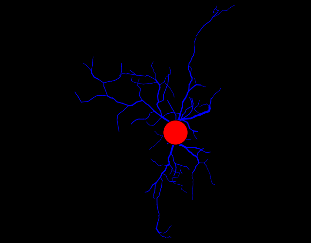
| NeuroMorpho.Org ID : | NMO_90195 |
| Cell Name : | CONT-2-_11 |
| Archive Name : | Diniz |
| Species Name : | mouse |
| Strain : | Albino |
| Structural Domains : | Processes, Soma |
| Physical Integrity : | Processes Moderate |
| Morphological Attributes : | Diameter, 3D, Angles |
| Min Age : | 16.0 months
|
| Max Age : | 20.0 months
|
| Gender : |
Female
|
| Min Weight : | Not reported
|
| Max Weight : | Not reported
|
| Development : | old |
| Primary Brain Region : | hippocampus |
| Secondary Brain Region : | CA3 |
| Tertiary Brain Region : | Not reported |
| Primary Cell Class : | Glia |
| Secondary Cell Class : | microglia |
| Tertiary Cell Class : | Iba1-positive |
| Original Format : | Neurolucida.asc |
| Experiment Protocol : | in vitro |
| Experimental Condition : | Piry virus-infected |
| Staining Method : | immunostaining |
| Slicing Direction : | Not reported |
| Slice Thickness : | 70
μm |
| Tissue Shrinkage : |
Reported 75% in z
Corrected 75%
|
| Objective Type : | oil |
| Magnification : | 100x |
| Reconstruction Method : | Neurolucida |
| Date of Deposition : | 2017-12-12 |
| Date of Upload : | 2018-08-02 |
| Persistence Vector : | CONT-2-_11.pvec |
|
|
| Reference Article |
| Related Article Reference : | Three-dimensional morphometric analysis of microglial changes in a mouse model of virus encephalitis: age and environmental influences. |
|
|
|
|
| Measurements |
| Soma Surface : |
158.12 μm2 |
| Number of Stems : |
7 |
| Number of Bifurcations : |
42 |
| Number of Branches : |
91 |
| Overall Width : |
57.87 μm |
| Overall Height : |
38.91 μm |
| Overall Depth : |
3.12 μm |
| Average Diameter : |
0.55 μm |
| Total Length : |
449.81 μm |
| Total Surface : |
824.24 μm2 |
| Total Volume : |
157.69 μm3 |
| Max Euclidean Distance : |
40.54 μm |
| Max Path Distance : |
45.64 μm |
| Max Branch Order : |
12 |
| Average Contraction : |
0.93 |
| Total Fragmentation : |
497 |
| Partition Asymmetry : |
0.58 |
| Average Rall's Ratio : |
1.3 |
| Average Bifurcation Angle Local : |
72.08° |
| Average Bifurcation Angle Remote : |
70.69° |
| Fractal Dimension : |
1.04 |
|
|
|
|
|
 0
0