|
| Details about selected cell |
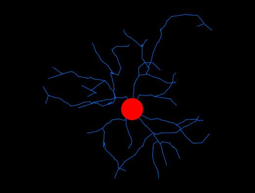
| NeuroMorpho.Org ID : | NMO_250046 |
| Cell Name : | A4Control_1 |
| Archive Name : | Garcia-Hernandez_DeSantis |
| Species Name : | rat |
| Strain : | Wistar |
| Structural Domains : | Processes, Soma |
| Physical Integrity : | Processes Moderate |
| Morphological Attributes : | No Diameter, 3D, Angles |
| Min Age : | 2.0 months
|
| Max Age : | 3.0 months
|
| Gender : |
Male
|
| Min Weight : | 300 grams
|
| Max Weight : | 400 grams
|
| Development : | adult |
| Primary Brain Region : | hippocampus |
| Secondary Brain Region : | dentate gyrus |
| Tertiary Brain Region : | granule layer |
| Primary Cell Class : | Glia |
| Secondary Cell Class : | microglia |
| Tertiary Cell Class : | Iba1-positive |
| Original Format : | Neurolucida.dat |
| Experiment Protocol : | in vivo |
| Experimental Condition : | Control |
| Staining Method : | immunostaining |
| Slicing Direction : | coronal |
| Slice Thickness : | 50
μm |
| Tissue Shrinkage : |
Not reported
|
| Objective Type : | dry |
| Magnification : | 20x , 40x |
| Reconstruction Method : | Neurolucida |
| Date of Deposition : | 2022-11-02 |
| Date of Upload : | 2022-12-07 |
| Persistence Vector : | A4Control_1.pvec |
|
|
| Reference Article |
| Related Article Reference : | Mapping microglia and astrocyte activation in vivo using diffusion MRI. |
|
|
|
|
| Measurements |
| Soma Surface : |
210.75 μm2 |
| Number of Stems : |
6 |
| Number of Bifurcations : |
22 |
| Number of Branches : |
50 |
| Overall Width : |
56.62 μm |
| Overall Height : |
57.6 μm |
| Overall Depth : |
3.06 μm |
| Average Diameter : |
0.25 μm |
| Total Length : |
499.66 μm |
| Total Surface : |
392.43 μm2 |
| Total Volume : |
24.53 μm3 |
| Max Euclidean Distance : |
42.59 μm |
| Max Path Distance : |
59.46 μm |
| Max Branch Order : |
6 |
| Average Contraction : |
0.91 |
| Total Fragmentation : |
335 |
| Partition Asymmetry : |
0.44 |
| Average Rall's Ratio : |
2 |
| Average Bifurcation Angle Local : |
91.55° |
| Average Bifurcation Angle Remote : |
78.77° |
| Fractal Dimension : |
1.04 |
|
|
|
|
|
 0
0