|
| Details about selected cell |
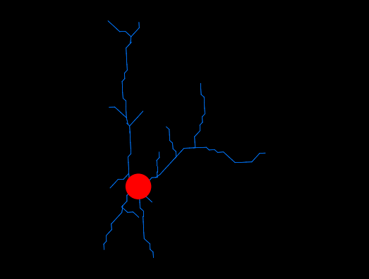
| NeuroMorpho.Org ID : | NMO_168075 |
| Cell Name : | 7828-day1-isoflurane-1-SR-T100_c |
| Archive Name : | Suzuki_Dityatev |
| Species Name : | mouse |
| Strain : | CX3CR1-EGFP x C57BL/6J |
| Structural Domains : | Processes, Soma |
| Physical Integrity : | Processes Moderate |
| Morphological Attributes : | No Diameter, 2D, Angles |
| Min Age : | 3.0 months
|
| Max Age : | 4.0 months
|
| Gender : |
Male
|
| Min Weight : | Not reported
|
| Max Weight : | Not reported
|
| Development : | adult |
| Primary Brain Region : | neocortex |
| Secondary Brain Region : | Not reported |
| Tertiary Brain Region : | Not reported |
| Primary Cell Class : | Glia |
| Secondary Cell Class : | microglia |
| Tertiary Cell Class : | C-X3-C Motif Chemokine Receptor 1-positive |
| Original Format : | Simple Neurite Tracer.swc |
| Experiment Protocol : | in vivo |
| Experimental Condition : | acute implant, isoflurane-anesthetized |
| Staining Method : | green fluorescent protein |
| Slicing Direction : | Sagittal |
| Slice Thickness : | Not reported
|
| Tissue Shrinkage : |
Not reported
|
| Objective Type : | water |
| Magnification : | 20x |
| Reconstruction Method : | Simple Neurite Tracer |
| Date of Deposition : | 2019-07-31 |
| Date of Upload : | 2021-11-16 |
| Persistence Vector : | 7828-day1-isoflurane-1-SR-T100_c.pvec |
| Note : | This reconstruction was obtained 1 day after surgery (quadrant 3). After imaging in awake mice, 2-h rest was given to each animal before imaging in the isoflurane-anesthetized condition. The same procedures of imaging as in awake-condition were applied in the other hemisphere, but the animal was head-fixed using a custom-made frame with a heating pad to keep body temperature constant, and anesthetized by 1.5% isoflurane during the imaging, which started 10 min after anesthesia induction. |
|
|
| Reference Article |
| Related Article Reference : | In vivo Two-Photon Imaging of Anesthesia-Specific Alterations in Microglial Surveillance and Photodamage-Directed Motility in Mouse Cortex. |
|
|
|
|
| Measurements |
| Soma Surface : |
194.83 μm2 |
| Number of Stems : |
4 |
| Number of Bifurcations : |
13 |
| Number of Branches : |
30 |
| Overall Width : |
67.87 μm |
| Overall Height : |
40.19 μm |
| Overall Depth : |
0 μm |
| Average Diameter : |
0.25 μm |
| Total Length : |
233.35 μm |
| Total Surface : |
183.27 μm2 |
| Total Volume : |
11.45 μm3 |
| Max Euclidean Distance : |
51.6 μm |
| Max Path Distance : |
58.44 μm |
| Max Branch Order : |
8 |
| Average Contraction : |
0.95 |
| Total Fragmentation : |
165 |
| Partition Asymmetry : |
0.69 |
| Average Rall's Ratio : |
2 |
| Average Bifurcation Angle Local : |
44.55° |
| Average Bifurcation Angle Remote : |
59.24° |
| Fractal Dimension : |
1.02 |
|
|
|
|
|
 0
0