|
| Details about selected cell |
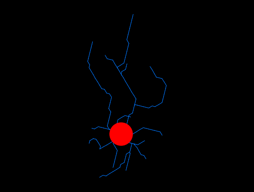
| NeuroMorpho.Org ID : | NMO_168060 |
| Cell Name : | 7828-day1-awake-SR-T100_c |
| Archive Name : | Suzuki_Dityatev |
| Species Name : | mouse |
| Strain : | CX3CR1-EGFP x C57BL/6J |
| Structural Domains : | Processes, Soma |
| Physical Integrity : | Processes Moderate |
| Morphological Attributes : | No Diameter, 2D, Angles |
| Min Age : | 7.0 months
|
| Max Age : | 8.0 months
|
| Gender : |
Male
|
| Min Weight : | Not reported
|
| Max Weight : | Not reported
|
| Development : | adult |
| Primary Brain Region : | neocortex |
| Secondary Brain Region : | Not reported |
| Tertiary Brain Region : | Not reported |
| Primary Cell Class : | Glia |
| Secondary Cell Class : | microglia |
| Tertiary Cell Class : | C-X3-C Motif Chemokine Receptor 1-positive |
| Original Format : | Simple Neurite Tracer.swc |
| Experiment Protocol : | in vivo |
| Experimental Condition : | chronic implant, awake |
| Staining Method : | green fluorescent protein |
| Slicing Direction : | Sagittal |
| Slice Thickness : | Not reported
|
| Tissue Shrinkage : |
Not reported
|
| Objective Type : | water |
| Magnification : | 20x |
| Reconstruction Method : | Simple Neurite Tracer |
| Date of Deposition : | 2019-07-31 |
| Date of Upload : | 2021-11-16 |
| Persistence Vector : | 7828-day1-awake-SR-T100_c.pvec |
| Note : | This reconstruction was obtained 4 months post-surgery (quadrant 1). FMice were head-fixed under the two-photon microscope using a Mobile HomeCage device in which nimals could freely move a light cage around them. After 10 min habituation, time-lapse imaging was performed for 33 min (100 frames, 20 s interval) by recording z-stacks (6 optical sections with a step size of 2 microns) around 100-150 microns below the pial surface to monitor microglial morphology and dynamics in the resting state. A single cell laser ablation was then achieved by focusing a two-photon laser beam at a single microglia cell in the superficial layer of the cortex to induce a highly precise and reproducible local injury. Immediately after, the same imaging procedure as in the resting state was repetead. |
|
|
| Reference Article |
| Related Article Reference : | In vivo Two-Photon Imaging of Anesthesia-Specific Alterations in Microglial Surveillance and Photodamage-Directed Motility in Mouse Cortex. |
|
|
|
|
| Measurements |
| Soma Surface : |
304.52 μm2 |
| Number of Stems : |
4 |
| Number of Bifurcations : |
10 |
| Number of Branches : |
24 |
| Overall Width : |
58 μm |
| Overall Height : |
31.04 μm |
| Overall Depth : |
2.99 μm |
| Average Diameter : |
0.25 μm |
| Total Length : |
275.3 μm |
| Total Surface : |
216.22 μm2 |
| Total Volume : |
13.51 μm3 |
| Max Euclidean Distance : |
50.7 μm |
| Max Path Distance : |
57.16 μm |
| Max Branch Order : |
4 |
| Average Contraction : |
0.9 |
| Total Fragmentation : |
195 |
| Partition Asymmetry : |
0.4 |
| Average Rall's Ratio : |
2 |
| Average Bifurcation Angle Local : |
24.31° |
| Average Bifurcation Angle Remote : |
70.59° |
| Fractal Dimension : |
1.04 |
|
|
|
|
|
 0
0